Revolutionary HEKA ElProScan Single-Cell Workstation Arrives at Hunan University of Science and Technology
The photoelectrochemical research team from the School of Chemistry and Chemical Engineering, led by Professor Chen Shu, Associate Professor Zhang Jie, and Associate Professor Liu Canjun, plan to integrate it with their research focused on single-particle electrochemistry, photo/electrocatalysis, electrochemical sensing, in vivo electroanalytical chemistry, and other biomedical application fields, making it an electrophysiology-electrochemistry multi-function combined platform.
Hunan University of Science and Technology has taken a significant leap forward in advanced research capabilities with the successful delivery and installation of the HEKA ElProScan Single-Cell Electrophysiology and Electrochemistry Nano-imaging Workstation. This high-precision, super-all-in-one system, custom-built by HEKA Elektronik (an affiliate of Harvard Bioscience), was put into use at the university's Analysis and Testing Center.
The implementation of the ElProScan system marks a pivotal moment, as it enables multi-scale imaging and analysis of the electrochemical activity and surface morphology of subcellular structures and nanomaterial surfaces. This powerful cutting-edge tool provides unprecedented capabilities for multi-dimensional analysis, imaging, and high-throughput screening across vital research areas.
Specifically, the system will be a cornerstone for advancements in discovering new energy materials and devices and neuroscience research. The photoelectrochemical research team from the School of Chemistry and Chemical Engineering, led by Professor Chen Shu, Associate Professor Zhang Jie, and Associate Professor Liu Canjun, will manage the device. They plan to integrate it with their research focused on single-particle electrochemistry, photo/electrocatalysis, electrochemical sensing, in vivo electroanalytical chemistry, and other biomedical application fields, making it an electrophysiology-electrochemistry multi-function combined platform.
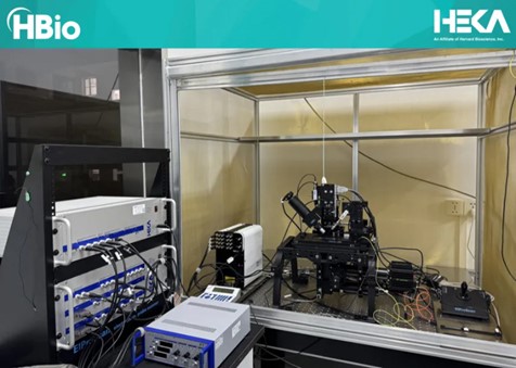
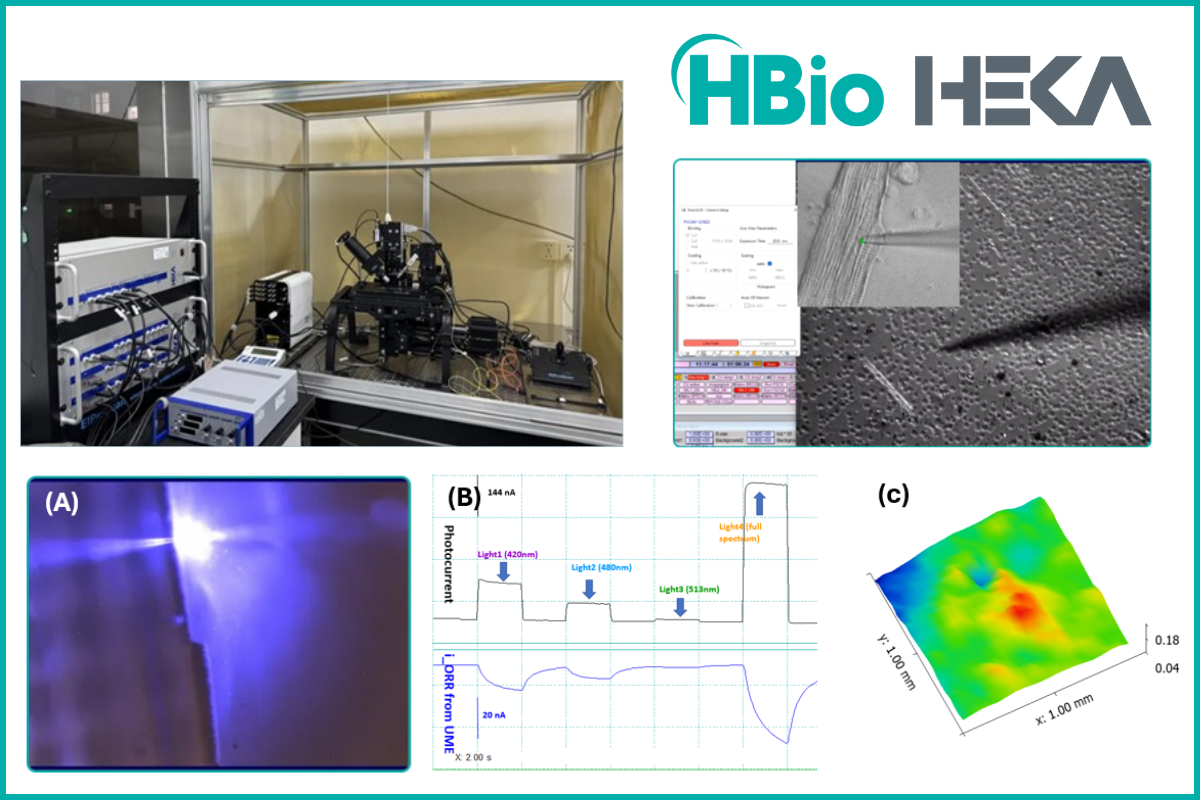
Figure 1. ElProScan single-cell electrophysiology and electrochemistry nano-imaging workstation.
Key Capabilities and Integrated Technologies:
The ElProScan workstation is a unique powerhouse due to its integration of several advanced technologies:
Integrated Electrochemical Scanning Probe Microscopy (ESPM) Technologies:
The system combines four major ESPM imaging techniques:
- SECM (scanning electrochemical microscopy)
- SECCM (scanning electrochemical cell microscopy)
- SICM (scanning ion conductance microscopy)
- SPECM (scanning photoelectrochemical microscopy)
These high-resolution scanning probe techniques allow researchers to analyze and map the electrochemical activity of micro/nano-material surfaces, including topography, redox activity, ion migration and conductance properties, localized electrochemical impedance, and interface capacitance. Crucially, it can correlate this E-chem activity with high-resolution three-dimensional morphology imaging captured in situ under photoelectrochemical reactions.
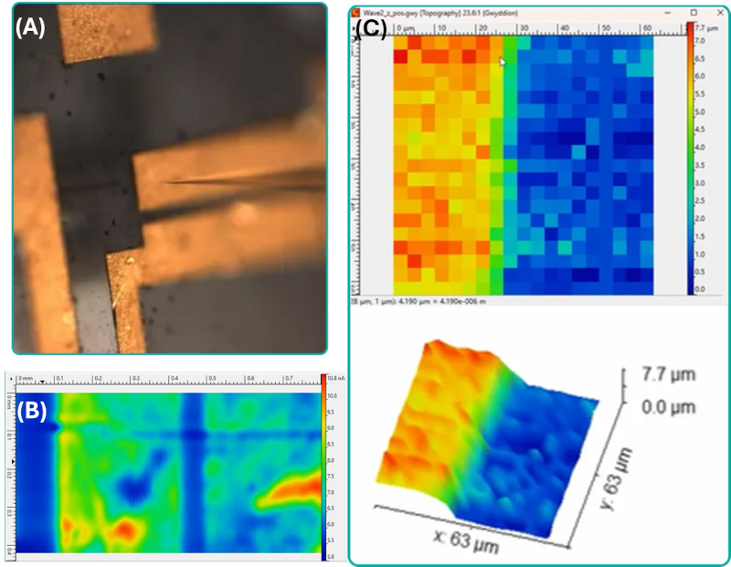
Figure 2. (A) Tip-to-surface prepositioning photo taken from top-45° imaging system; (B) electrochemical activity map of a metal coating recorded in SECM mode; (C) 3D topography map of metal surface coating recorded in situ via SECM probe by HEKA’s Shear-force sensing technology.
Spatially Resolved Photoelectrochemical Imaging:
Utilizing HEKA's unique modular dual light-path inverted microscope design, the system enables spatially resolvable scanning photoelectrochemical microscopy, by virtual of its flexible adjustment of the incident light beam spot-size and the incident light-path directions. The SPECM technology, powered with a multi-wavelength excitation light source (with 16-LED wavelength from 365nm to 770nm), allows for precise synchronized excitation/detection applications, such as:
- Imaging and analyzing local surface defects and active hot spots of semiconducting photocatalyst materials
- Imaging of micro-/nano-scale photoelectrocatalytic reaction rate and quantum efficiency (QE)
- Diversified photoelectrochemical (PEC) applications like solar-driven water-splitting and pollutant degradation in situ.
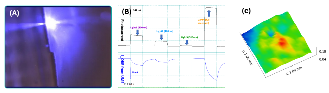
Figure 3. (A) Photograph showing precise alignment of microelectrode probe with colocalized tunable incident light beam spot; (B) synchronized dual-channel photocurrent and oxygen reduction current signals recorded at local photocatalysts coating in SPECM mode; (C) 3D map of water-splitting IPCE (Incident Photon-to-Electron Conversion Efficiency) of a semiconducting photocatalyst material.
Fully Functional Patch Clamp System:
Designed for imaging single cells at the micro- and nanoscale, the system features HEKA's EPC10USB double-channel patch clamp amplifiers and a customized micromanipulation system. This integration provides capabilities for intracellular and extracellular imaging, screening and sensing, including:
- Single cell precision microinjection and drug activity screening
- Whole-cell/single-channel patch clamp recordings
- Intercellular and intracellular neurotransmitter detection by FSCV (Fast-Scan Cyclic Voltammetry)
- Solid-state nanopore applications
- Nano-electrochemical confined detection based on nanoprobes
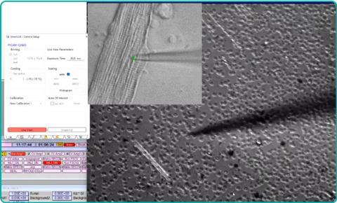
Figure 4. Optical photograph of a patch clamp micropipette tip that’s positioned under precise micromanipulation control (photo insert is a higher magnification image of a modified bifunctional sensor probe positioned over living cell surface).
In situ Synchronized Fluorescence Microscopy Imaging System:
A multi-angle optical synchronous monitoring module provides full-view visualization of the scanning probe operation process. Through a modular optical imaging design (45° oblique digital camera + inverted fluorescence microscope with dual-layer optical train + high-speed CCD camera system), the optical imaging system achieves fully automatic synchronous high-speed fluorescence imaging functions, supporting the most challenging ratiometric fluorescence imaging as well.
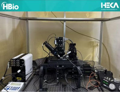
Figure 5. ElProScan electrophysiology and electrochemistry nano-imaging workstation - 4D XYZF Nanopositioning system with inverted modular synchronous fluorescence microscope
Importantly, the system ensures that synchronous fluorescence imaging data and all electrophysiological and electrochemical recording data are synchronously linked and saved inside one piece of HEKA Patchmaster software. This allows for flexible online and offline analysis, processing, and export of data.
Furthermore, the system supports customized ECL (electrochemiluminescence) imaging, easy integration of a micro-spectrophotometer, in situ integrated microfluidic systems, and customized MEA (multi-electrode array) system for high-throughput screening applications.
The arrival of the HEKA ElProScan workstation at Hunan University of Science and Technology signifies a major upgrade in its research infrastructure, promising to accelerate discoveries in materials science, energy-related photoelectrocatalysis, neuroscience and biomedical research fields by providing unparalleled insights into electrochemical and electrophysiological processes at the micro- and nanoscale.
Learn more about HEKA’s electrochemistry and patch clamp electrophysiology solutions, or contact us to discover how it can elevate your research.