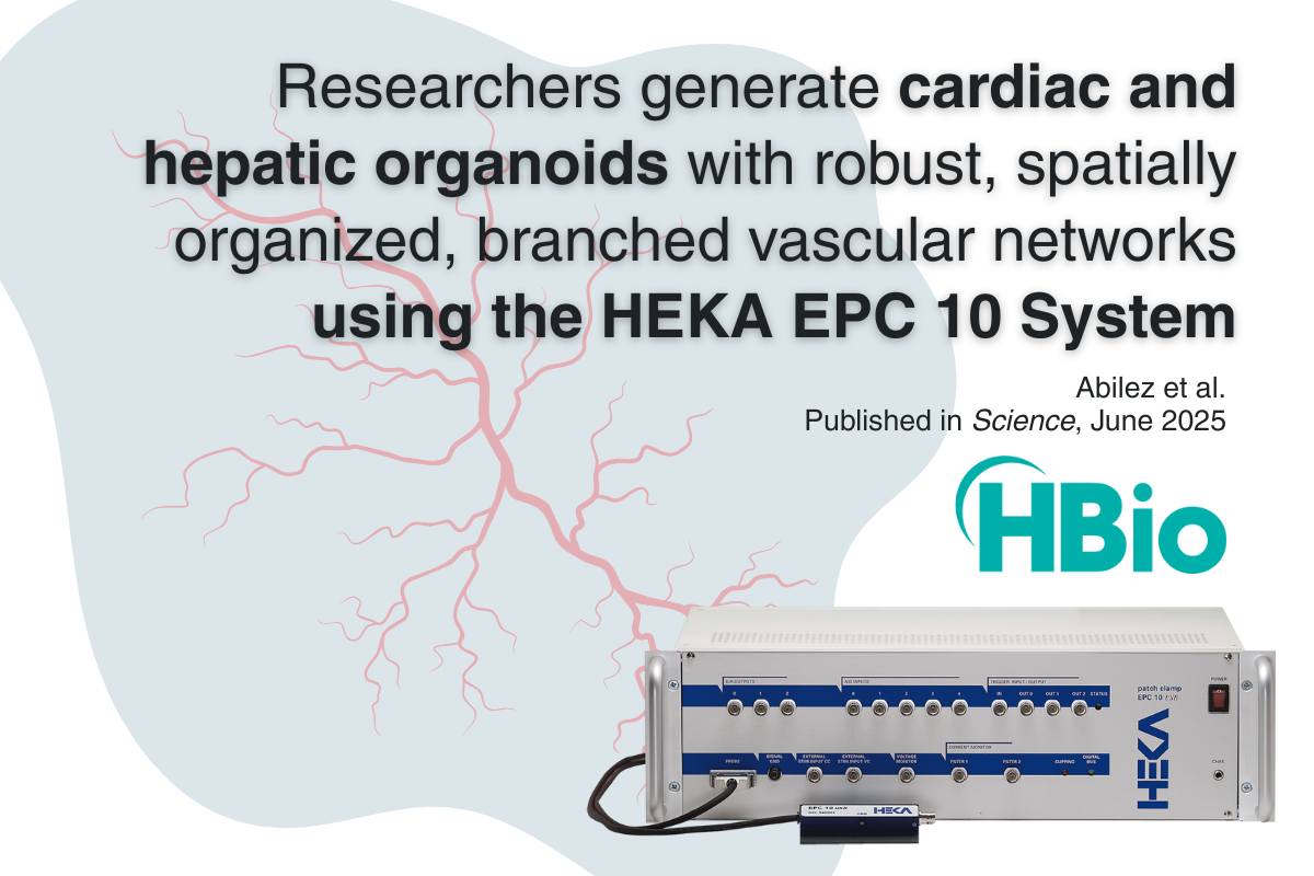
Vascularized Cardiac and Hepatic Organoids: A Leap Forward for Organoid Electrophysiology
In the Abilez et al. study, the HEKA EPC 10 Patch Clamp Amplifier provided the sensitivity, stability, and control needed to investigate individual cells within the complex 3D environment of the organoid.
A new study from Abilez et al., published in Science (June 2025), represents a major advance in our ability to model human organ development in vitro. By combining geometric micropatterning of human pluripotent stem cells (hPSCs) with a carefully optimized “vascular-inducing cocktail” of growth factors, the team successfully generated cardiac and hepatic organoids with robust, spatially organized, branched vascular networks, complete with lumina and integrated with multiple relevant cell types.
This breakthrough matters for developmental biology, disease modeling, and regenerative medicine – but it also carries big implications for organoid electrophysiology.
Why Vascularization Matters for Electrophysiology
Electrophysiological recording, whether patch clamp or multielectrode array, depends on having physiologically active, viable cells deep within the tissue. In conventional non-vascularized organoids, diffusion limits mean that cells in the core can become hypoxic, necrotic, or electrically silent. By establishing a branched vascular system early in development, these cardiac vascularized organoids (cVOs) and hepatic vascularized organoids (hVOs) maintain healthier, more electrically representative cell populations throughout their 3D volume.
For researchers, this means:
- Higher probability of viable target cells in the interior of an organoid.
- Improved functional maturity – vascularization correlates with more physiologically relevant ion channel expression.
- Better reproducibility in electrophysiological measurements, thanks to reduced heterogeneity caused by necrotic zones.
Patch Clamp Precision with the HEKA EPC 10 Patch Clamp Amplifier System
Capturing the fine details of how cells in these vascularized organoids behave electrically requires equipment capable of measuring extremely small currents with minimal noise. In the Abilez et al. study, the HEKA EPC 10 Patch Clamp Amplifier provided the sensitivity, stability, and control needed to investigate individual cells within the complex 3D environment of the organoid. Its precision allowed researchers to characterize the electrophysiological properties of different cell types and confirm functional integration within the newly formed vascular networks, an essential step in validating that these organoids mimic early human cardiac and hepatic development.
In 2025, HEKA introduced an updated version of the EPC 10 system, the EPC 10 USB 3.0 Patch Clamp Amplifier, featuring new capabilities, refinements, and software improvements.
The Planar MEA Question for 3D Organoids
Multielectrode arrays (MEAs) are a powerful complement to patch clamp, offering long-term, network-level recordings. However, it’s worth reflecting on the geometry. Planar (2D) MEAs – like those used in many commercial systems – place electrodes on a flat surface. This works beautifully for monolayers and thin slices, but in a spherical, millimeter-scale 3D organoid, most of the active cells are not in direct contact with the electrodes. Signals from deep layers are attenuated, and the picture you get is biased toward surface activity.
This doesn’t mean planar MEAs are “wrong” for organoids, but it’s food for thought: if your model is inherently 3D, shouldn't your recording approach be as well?
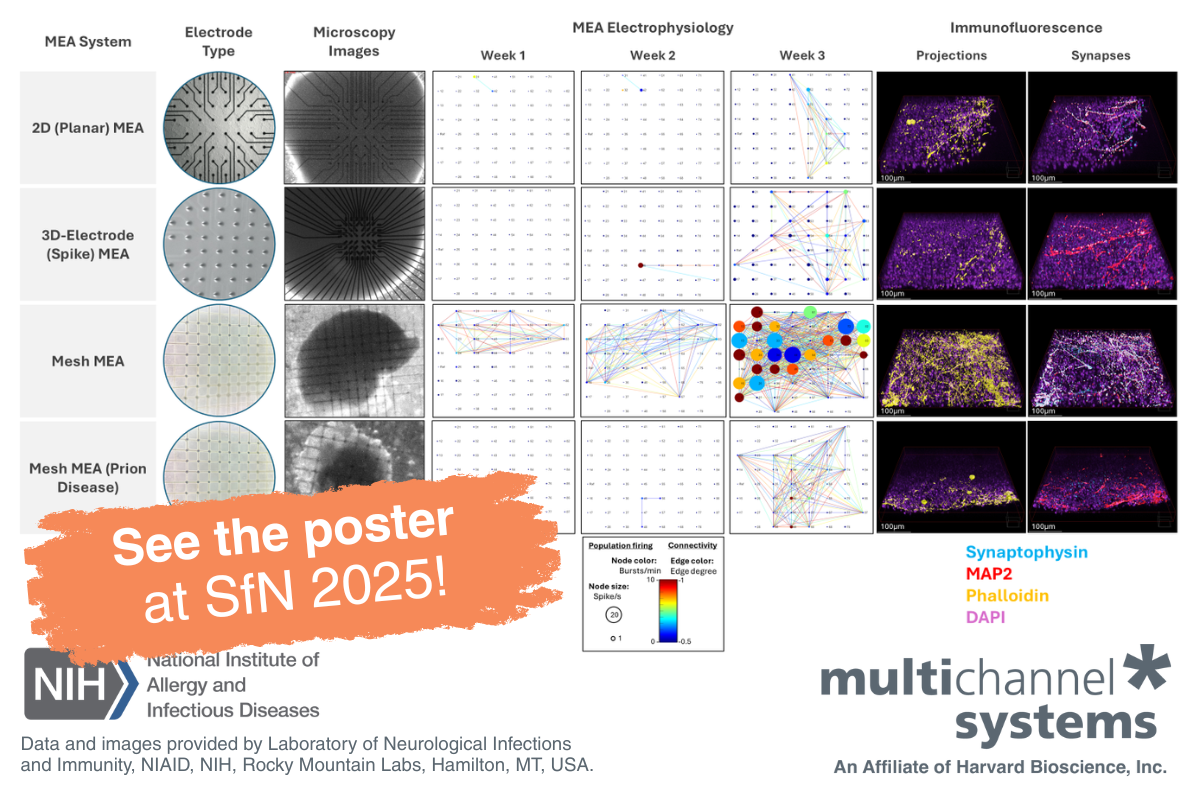
A 3D-Optimized Alternative: Mesh MEA from Multi Channel Systems
For researchers seeking full-volume coverage of organoid activity, Mesh MEA from Multi Channel Systems provides a solution designed specifically for 3D cultures.
- Internal MEA recordings: the mesh structure is embedded within the organoid, capturing signals from its entire depth.
- Chronic recordings: monitor the same organoid for days or weeks without disrupting growth.
- Improved spatial resolution: record from multiple layers simultaneously to map network development.
- Compatibility with perfusion and imaging, supporting multimodal experiments.
- Proven for organoid and spheroid models: validated in neuronal and cardiac applications.
By pairing advanced biological models like the vascularized cVOs and hVOs from this study with recording systems that match their complexity, researchers can open new frontiers in developmental electrophysiology, disease modeling, and drug testing.
Paint a fuller picture of organoid activity by pairing Mesh MEA’s long-term, network-wide insights with the HEKA EPC 10 USB 3.0’s Patch Clamp Amplifier’s unrivaled single-cell precision.
Learn more about the EPC 10 USB 3.0 Patch Clamp Amplifier System.